pain management Archives

Three Types of Mast Cell Activation Syndrome

Mast cells are important cells in our immune systems that help us fight off sickness, infections, and allergies. Although these cells are vital to protecting our bodies, some peoples’ mast cells can be too active for no good reason. In these cases, the mast cells might cause more harm than good. When Mast Cells are overly active, Mast Cell Activation Syndrome (MCAS) may sometimes be diagnosed. In MCAS, Mast Cells release excessive chemicals into the bloodstream. This reaction can cause swelling, hives, coughing, and pain.

There are three main types of MCAS that are recognized:

  1. Primary
  2. Secondary
  3. Idiopathic.

Primary MCAS is when the bodies’ mast cells clone themselves excessively. In these cases, mast cells gather in organ tissues in excessive numbers. Symptoms of primary MCAS can occur in pretty much any organ in the body. Common symptoms are allergy-like. For example, skin rashes, hives, and abdominal pain. Especially serious cases of primary MCAS can be called mastocytosis, a life-threatening condition that can cause serious allergic reactions called anaphylaxis.


Even though you may have not heard of it, you are probably familiar with secondary MCAS. Do you have any allergies? If so, you may have secondary MCAS. Secondary MCAS may be diagnosed when a person experiences allergic reactions, for example, to nuts or shellfish. Unlike in primary MCAS, mast cells do not clone themselves abnormally in second MCAS. In fact, people with secondary MCAS have a perfectly normal amount of mast cells. However, these mast cells are overactive and release excessive chemicals into the bloodstream (such as histamine), which cause allergic like reactions. Some examples of environmental triggers can include pollen, food, and alcohol.


Idiopathic MCAS is an interesting condition that can be frustrating for patients who have it. This is because these patients experience allergy-like symptoms, but they are not allergic to anything. In other words, idiopathic MCAS can cause seemingly allergic reactions for no reason at all. For MCAS to fall into the idiopathic category, there must be no evidence of an excessive amount of mast cells (primary MCAS), and the allergy-like symptoms must not be triggered by an actual allergy. For someone to be diagnosed with idiopathic MCAS, the doctor must make sure that they rule out all potential causes of the allergy symptoms.  Although not typically, patients with idiopathic MCAS may still have serious reactions (anaphylaxis).MCAS in all its forms is treatable through different strategies. Helpful interventions for primary, secondary and idiopathic MCAS may include antihistamines to calm allergy symptoms. For cases of secondary MCAS where the allergic trigger is known, lifestyle changes can go a long way in calming down mast cells.


Share

Idiopathic Mast Cell Activation Syndrome (MCAS)

Are your “allergic symptoms” and pain related to idiopathic Mast Cell Activation Syndrome (MCAS)?

Hives, rashes, itching, problems breathing, and pain in your stomach or other parts of your body may all be related to allergies. Your doctor may have tested you, but did not find what you might be specifically allergic to, for example types of foods or substances surrounding us like pollen, dust, and mold. If you didn’t have evidence of an allergy to any or only a few of these substances, then what is causing your symptoms?

Your symptoms may be caused by excessive activity in a special cell in your body called the Mast Cell (MC) which is stimulated whenever you experience any irritation. The MC helps to fight off infections and help repair damaged tissue by producing many different powerful substances. However, when it is overactive, these substances may be produced by the MC after exposure to ordinary stimuli including a hot shower, sunlight, perfumes, cleaning solutions, and a variety of foods. Overactive MCs may cause diffuse pain in various parts of your body.

Doctors have been aware that increased numbers of MCs can cause serious symptoms in different organs. When there are too many MCs the diagnosis is often Mastocytosis, which requires specialized treatment. When the number of MCs is normal and the MCs are just overactive, the diagnosis, which is a relatively new discovery, is often idiopathic Mast Cell Activation Syndrome (MCAS) and can be diagnosed with several blood and urine tests. However, if you have symptoms in 2 or more organ systems, for example skin rashes, heart burn, asthma like symptoms, and your symptoms improve with an anti-histamine, you may be diagnosed with idiopathic MCAS without complicated testing.

Our body makes a substance that can down-regulate the activity of the MC. It is called palmitoylethanolamide (PEA). It is in the family of substances, called cannabinoids, and is related to the molecules that are present in marijuana. Other substances can down regulate the MC such as Cromolyn Sodium. Since one of the main substances produced by the MC is histamine, medications that block histamine. such as antihistamines used for allergies and histamine antagonists used for heart burn, may be helpful in reducing the symptoms of overactive MCs.

If you have diffuse pain and other symptoms, like the ones above, that have been difficult to diagnose and treat, you may have MC problems that can be treated.


Share

What is a clinical trial

How do we test new medicines before they are released to the public?

Let’s say for the sake of this example that there’s a new drug that is potentially effective at treating people with muscle pain. The process to test that treatment is called clinical trials. To test the medicine, clinical trials occur in different stages, called phases. Phase I testing is done on healthy volunteers without muscle pain. If the drug is found have unsafe side effects, it would fail phase 1 and be withdrawn from further testing. If the drug passes Phase 1, it goes on to Phase 2 which studies a small group of patients with muscle pain to see if the new drug is effective in treating the condition without producing serious side effects. If the drug passes Phase 2 it goes on to Phase 3 which tests a larger group of patients with muscle pain to confirm the effectiveness and safety found in Phase 2.

There are strict guidelines that researchers must follow when conducting clinical trials to ensure the safety of their human subjects. Regulatory bodies such as the FDA monitor clinical trials to ensure that all guidelines are being followed. If the physicians in charge of a new drug trial are found to not follow the guidelines, they are removed from their trial and may lose their ability to participate in any future trials.

Participation in a new drug study can be an exciting experience–especially when it results in a decrease or elimination of your symptoms. Before a patient begins a clinical trial, all the possible risks that may occur during the study are spelled out in a document called the consent form. The consent form is signed by the participant before they start treatment. The consent form makes it clear that declining to participate in a study will not affect the participant’s future care, and that they may withdraw from the study at any time for any reason.

A drug study is an experiment. To ensure that enthusiasm about the new drug doesn’t influence the outcome–for example hoping that the treatment is going to work will make you believe that it works–some participants will receive an inactive drug called a placebo. Whenever possible, the physician administering the drug will not know if they are giving you the real drug or the placebo. This type of study, where the physician and the study participant do not know which medication they receive, is called a double-blind study. Participants are informed at the end of the study if they received the active drug or the placebo.

Some drug trials provide financial compensation for time and participation. Some trials will only compensate you for any expenses that you incur in the process of participating in the study. Participation in a study may or may not benefit you directly but will potentially benefit many patients in need of new effective treatments.

Share

Sitting with your legs crossed may be contributing to your back pain

Think of some of the ways you sit. Whether taking a ride on the subway, waiting patiently at the doctor’s office, or sitting as a passenger in a car, the way we sit, especially for prolonged periods can have an effect on the pain we may experience in our low back.


For many of us, sitting with our legs crossed feels comfortable in the moment but have you ever considered how this can affect your back and cause pain? What happens to your spine and muscles when sitting with your legs crossed for an extended period of time? When considering good posture, a large component is keeping your spine neutral and avoiding positions that twist the spine or cause misalignment. When sitting with your legs crossed, your pelvis becomes tilted and unconsciously, we tilt our entire torso towards one side. There is a curvature towards one side in the low back which then creates problems in other areas of the spine. Naturally, with a tilt of the pelvis, the upper body will compensate by leaning the opposite direction to maintain balance. Working from the bottom up, pressure on the lumbar spine will cause curvature and misalignment in the thoracic spine. This places strain on the cervical spine to keep the head in the upright position.


Sitting with our legs crossed can also affect our muscles. There are two common sitting positions that we should be mindful of, knee over knee or foot over knee for a prolonged period can cause a tightness in the hamstrings, hip flexors and glute muscles. The iliopsoas, one of the major hip flexor muscles, is responsible for external rotation of the femur and an integral part in maintaining posture. This muscle may become strained due to contraction while the pelvis is tilted. You may notice soreness and tenderness in the low back and hip area. Similarly, sitting foot over knee can over time damage the muscles in the inner thigh. Most commonly, the sartorius muscle can become tight and cause discomfort when sitting with the calf over the knee. At the Norman Marcus Pain Institute, Iliacus and Psoas muscles on the hip can be treated for pain. As one of the most common muscles causing pain for patients, when treated, the Iliacus and other hip flexors can provide great relief for pain in the low back, buttock and groin.

Instead of sitting with crossed legs, sit in a chair with height necessary to place both feet flat on the floor. While sitting upright with your back against the chair, you may want to consider placing a cushion under your bottom that can add support to the low back.

References

Lee BJ, Cha HG, Lee WH. The effects of sitting with the right leg crossed on the trunk length and pelvic torsion of healthy individuals. J Phys Ther Sci. 2016 Nov;28(11):3162-3164. doi: 10.1589/jpts.28.3162. Epub 2016 Nov 29. PMID: 27942141; PMCID: PMC5140821.

Share

Best Posture for Reading: How to alleviate back pain

There’s nothing better than snuggling into a good book. But are you experiencing back pain while sitting and reading for an extended period? Back pain can often be caused by improper posture. While reading, whether using a physical book or an electronic device, many people experience back pain in the neck and shoulder or in the low back. Here are some ways to alleviate back pain while reading.

Reading can cause strain on the neck and shoulders if there is a tilt in the neck to look down at your book or device. While sitting and reading, it’s important to place your reading material at eye level. Sitting with your head leaning forward and back hunched can cause hyperflexion of the cervical spine and lead to severe neck and back pain over time. It is best to use a stand that can be adjusted to the required height. Many have recommended raising your materials with your hand; however, this may also cause some strain on the neck and shoulders. While holding an object in front of you at eye level, either with one or both hands, your shoulder and neck muscles are in constant contraction to sustain this positioning. Even over a period of a few minutes, this can cause the muscles to spasm or cause intense strain. Using an adjustable stand will ensure that we can sit straight up, taking pressure off of the cervical spine.

The best posture for reading is sitting upright in a chair with lumbar support. Avoid sitting on a seat that lacks back support such as a stool or a bench. A chair with good ergonomics is one that supports the low back and provides an arm rest to place both elbows. Mentioned above, we want to avoid stiffness in the upper back and neck muscles. Placing your elbows on an arm rest that is low enough to support your arms without adding additional strain is ideal. A study examining the association of low back pain with cell phone use found that thoracolumbar kyphosis and lumbar lordosis (curvature in the spine causing a pelvis tilt) increased with prolonged sitting. Participants had a slouch and progressed spine curvature after sitting for longer than 30 mins. The study also found that those with pre-existing back pain had a significantly higher increase in lordosis and complained of more back pain. When sitting for longer than 30 minutes, take breaks. Sitting should be interrupted by standing breaks to keep blood flowing and reduce stiffness in muscles.

To summarize, here are some things we can do to minimize back pain while reading or using a device while sitting.

– Sit with back and arm support for your neck and shoulders

– Bring your reading material or device to eye level to avoid neck strain

– Take breaks and stand for a minute or so while sitting for more than 30 minutes

References

In TS, Jung JH, Jung KS, Cho HY. Spinal and Pelvic Alignment of Sitting Posture Associated with Smartphone Use in Adolescents with Low Back Pain. Int J Environ Res Public Health. 2021 Aug 7;18(16):8369. doi: 10.3390/ijerph18168369. PMID: 34444119; PMCID: PMC8391723.

Share

How your sleeping position affects your back pain and neck pain

We all have different preferences when it comes to sleeping. Some of us are side sleepers while others may prefer to sleep on our backs. But there may be a few things to consider about your sleeping position that may be contributing to your back pain. Here are three ways to sleep to help prevent back pain and get a good night’s rest.

Sleeping on your side.

When laying on your side. Be sure to first support your head and neck. This can be done by placing as many pillows or supports as needed to keep the cervical spine neutral and the muscles surrounding it relaxed. These pillows or supports should be placed in the gap between the neck and the head, until the head is upright. Try to avoid raising your arm above your head as this may cause additional strain in the neck and shoulders. To keep the lumbar spine as neutral as possible, place a pillow between your knees. You may need to find a pillow that has enough support to hold the leg. This prevents the hips from rotating forward toward whichever side you are leaning on and helps to prevent rotation in the hips.

Sleeping on your back.

The recommendations for sleeping will be the same with slight adjustments for positioning. You must support your head and neck while sleeping on your back. This can be accomplished by placing your pillows in the gap between your neck and the bed while ensuring the top of the head is also lifted. This prevents an over extension of the cervical spine. Secondly, place a pillow under your knees to raise them slightly above the pelvis. This tilts the sacrum and the spine to neutral position.

Sleeping on your stomach/front.

A study on the relationship between sleep posture and spinal symptoms found that the prone sleeping position or sleeping on your front is the largest contributor back pain and poor quality of sleep. Although this sleeping position is not recommended for extended periods of time. If you need to sleep on your front, place a pillow under your hips to prevent curvature in the lower back. Additionally, a pillow should be placed under the ankles to provide comfort for the knees and avoid hyper extension of the hamstrings. Although it may be comfortable in the moment, raising a knee to either side may cause additional rotation in the sacrum and over time cause strain on the hip flexor muscles and muscles surrounding the spine.

To summarize, here are some ways you can avoid back pain while sleeping:

– Support your head and neck

– Avoid curvature in the back and rotation of sacrum for an extended period of time.

– Use pillows when necessary for additional comfort.

References

Cary D, Briffa K, McKenna L. Identifying relationships between sleep posture and non-specific spinal symptoms in adults: A scoping review. BMJ Open. 2019 Jun 28;9(6):e027633. doi: 10.1136/bmjopen-2018-027633. PMID: 31256029; PMCID: PMC6609073.

Cary D, Jacques A, Briffa K. Examining relationships between sleep posture, waking spinal symptoms and quality of sleep: A cross sectional study. PLoS One. 2021 Nov 30;16(11):e0260582. doi: 10.1371/journal.pone.0260582. PMID: 34847195; PMCID: PMC8631621.

Share

How neck pain affects your walking

A recent study done in Turkey took a look at the walking patterns of chronic pain patients- ideopathic neck pain, to be specific. They based their hypothesis around previous studies that have shown that people with chronic ideopathic neck pain (CINP) had abnormal walking patterns (known as gait), whether it be in the physical pattern or in the timing of their walks.

The scientists recruited 20 individuals with CINP as well as 20 healthy individuals without CINP to perform 10 minute walking tests with pressure sensitive insoles inserted into their shoes. They performed three different tests: Preferred walking, preferred walking with head rotation, and walking at maximum speed.

The study showed that individuals with CINP had a slower gait speed in all three walking conditions compared to the control groups. The gait was also found to be asymmetric in individuals with CINP in two out of three of the different test groups. This is important because gait asymmetry has the potential to load your entire body unequally, causing unbalanced strain on your muscles, joints, and other problem areas. These can lead to other musculoskeletal problems in the future.

Here at Norman Marcus Pain Institute, we see many patients with chronic pain everywhere, not just in the neck. We also often see collapsed arches in patients’ feet as well as uneven strides that can be magnifying discomfort on one side of the body vs the other. Recently we have started recommending special orthotics designed to strengthen and correct imbalances and weak arches. We occasionally recommend that our patients see a professional gait analyst to find ways to correct these imbalances to relieve some of the strain that their muscles may be experiencing.

Share

Ehlers-Danlos Syndrome (EDS) Demographics & Characteristics

A recent study released by Rheumatology takes a look at the demographics of 280 patients previously diagnosed with hypermobility syndrome. They gathered a cohort of 280 patients – 90% female and 10% male, between ages 18 and 66 (it is generally accepted that females have a higher rate of hypermobility than males, though it is not clear why). 95% of these patients had what was previously known as EDS type 3, now known as hypermobile Ehlers-Danlos Syndrome (hEDS). They proceeded to analyze a comprehensive medical history.

The average age of diagnosis of these patients was 29. 66% of patients had a family history of hypermobility, which corresponds with what we know about the genetic nature of Ehlers-Danlos syndrome. A family history of hypermobility and/or EDS is one of the accepted criteria to be diagnosed with hEDS.

66% of the patients had orthostatic intolerance, meaning their vitals changed dramatically when standing up and returned to “normal” when lying down. This includes drastic increases in heart rate and decreases in blood pressure due to the body’s inability to properly compensate for the challenges of standing upright. Almost half of those with orthostatic intolerance had bowel problems, compared to only 19% without intolerance. 45% had gastrointestinal problems, and 39% had bladder dysfunction. Chronic joint and muscle pain were reported in 91% of the patients. Approximately half of these patients with chronic pain used opioids long-term to relieve their pain. 43% patients reported being on work disability.

While the official clinical diagnoses for hypermobile EDS focus on joint hypermobility and skin hyperextensibility, the study confirms that this particular condition is systemic and also has a high correlation to orthostatic intolerance and chronic pain. Therefore it is important to look at a holistic, overall approach of each patient suspected to have hEDS in order to properly assess how to improve the quality of life of each person living with hEDS.

Ref: Dojcinovska, M., Cohen, H., & Wolman, R. (2019). 200 Demographics and clinical characteristics of a national cohort of 280 patients with hypermobility syndrome and impact on their work disability. Rheumatology, 58(Supplement_3), kez107-016.

Share

Updated Ehlers-Danlos Criteria

In May 2017, there were several changes in the criteria for the diagnosis of what used to be Ehlers-Danlos Syndrome Type III, hypermobility type. First, the name has been changed to hypermobile Ehler-Danlos Syndrome or hEDS. Instead of six types, there are now 13 types.


  Clinical EDS subtype Abbreviation IP Genetic Basis Protein
1 Classical EDS cEDS AD Major: COL5A1, COL5A1 Type V collagen
        Rare: COL1A1 Type I collagen
        c.934C>T, p.(Arg312Cys)  
2 Classical-like EDS clEDS AR TNXB Tenascin XB
3 Cardiac-valvular cvEDS AR COL1A2 (bilallelic mutations that lead to COL1A2 Type I collagen
        NMD and absence of pro α2(I) collagen chains)  
4 Vascular EDS vEDS AD Major: COL3A1 Type III collagen
        Rare: COL1A1 Type I collagen
        c.934C>T, p.(Arg312Cys)  
        c.1720C>T, p.(Arg574Cys)  
        c.3227C>T, p.(Arg 1093Cys)  
5 Hypermobile EDS hEDS AD Unknown Unknown
6 Arthrochalasia EDS aEDS AD COL1A1, COL1A2 Type I collagen
7 Dermatosparaxis EDS dEDS AR ADAMTS2 ADAMTS-2
8 Kyphoscoliotic EDS kEDS AR PLOD1 LH1
        FKBP14 FKBP22
9 Brittle Cornea Syndrome BCS AR ZNF469 ZNF469
        PRDM5 PRDM5
10 Spondylodysplastic EDS spEDS AR B4GALT7 β4GalT7
        B3GALT6 β3GalT6
        SLC39A13 ZIP13
11 Musculocontractural EDS mcEDS AR CHST14 D4ST1
        DSE DSE
12 Myopathic EDS mEDS AD or AR COL12A1 Type XII collagen
13 Periodontal EDS pEDS AD C1R C1r
        C1S C1s
           
IP, inheritance pattern; AD, autosomal dominant; AR, autosomal recessive; NMD, nonsence-mediated mRNA decay


hEDS continues to have no obvious known genetic testing and is only diagnosed through clinical testing.

As mentioned earlier, the Beighton scale is used to determine if a person is hypermobile. This continues to be an important determinant for hypermobility. A 5/9 score in adults is needed in order to meet this requisite. However, because age has an effect on flexibility (ie with age, you tend to become stiffer), there is a Five Point Questionnaire that determines if there is a history of hypermobility. The five points are the following:

  • Can you now (or could you ever) place your hands flat on the floor without bending your knees?
  • Can you now (or could you ever) bend your thumb to touch your forearm?
  • As a child, did you amuse your friends by contorting your body into strange shapes or could you do a split?
  • As a child or teenager, did your shoulder or kneecap dislocate on more than one occasion?
  • Did you consider yourself “double-jointed”?

If you answer yes to two or more questions, then you have met the criteria for hypermobility (with 80-85% sensitivity and 80-90% specificity).


The Beighton Scale and Five Point Questionnaire are the first part of three criteria that need to be met in order to qualify for the diagnosis of hEDS. This criterion also meets the diagnosis of hypermobility (you can have generalized joint hypermobility without a diagnosis of hEDS).


The second criterion has three parts (A, B, and C), of which two must be met to qualify. Part A is a series of 12 symptoms, of which five must be met in order to quality:

  • Unusually velvety skin
  • Mild skin hyperextensibility
  • Unexplained striae on back, groins, thighs, breast or abdomen in adolescents, men or prepubeteral women without a history of significant gain/loss or weight.
  • Bilateral piezogenic papules of the heel
  • Recurrent or multiple abdominal hernias (umbilical, inguinal, crural)
  • Atrophic scarring with papyraceous or meosideric scars
  • Pelvic floor, rectal and/or uterine prolapse in children, men or nulliparous women without a history of morbid obesity or other known predisposing medical conditions.
  • Dental crowding or high or narrow palate
  • Arachnodactyly (fingers & toes, long and slender), Steinberg Sign (thumb beyond palm), Walker-Murdoch sign (thumb overlapping fifth finger around wrist)
  • Arm span to height ≥ 1.05
  • MVP
  • Aortic root dilation


Part B is a positive family history of one or more first degree relatives, independently meeting the criteria of hEDS.

Part C is musculoskeletal complaints; a patient must have at least one to qualify:

  • Daily musculoskeletal pain at least three months in two or more limbs
  • Chronic widespread pain for more than 3 months.
  • Recurrent joint dislocation or frank instability in the absence of trauma (A or B)
    • 3 or more atraumatic joint dislocations in the same joint or two or more atraumatic dislocations in two different joints occurring at two different times.
    • Medical confirmation of joint (2 or more) instability


The third criterion contains three pre-requisites, all of which must be met:

  1. Absence of unusual skin fragility which should prompt consideration of other types of EDS.
  2. Exclusion of other heritable and acquired connective tissue disorder with autoimmune connective tissue disorder (ie Lupus, RA).
  3. Exclusion of alternative diagnoses that may also include joint hypermobility by means of hypotonia and/or CT laxity. Alternative diagnoses and diagnostic categories include, but not limited to, neuromuscular diseases, myopathic EDS, Bethlem myopathy, or HCTD (ie other types of EDS), Loeys-Dietz Syndrome (Marfan), and skeletal dysplasias (ie OI).


References

American Journal of Medical Genetics, Part C. (Seminars in Medical Genetics), March 2017.

Share

Ehlers-Danlos (EDS)

It is known that physically active patients who have stiff muscles frequently develop pain.  What is less known is that patients with overly flexible joints will also frequently develop muscle pain.

Congenital diseases, such as Marfan Syndrome and Ehlers-Danlos Syndrome (EDS), may cause joints to repeatedly dislocate, resulting in stiffness in the muscles surrounding the joint – a way that the body attempts to help the unstable joint.  The reflexive stiffness and spasms in the muscles surrounding the unstable joint cause pain.

Treatment to relax the muscles such as injections into trigger points or into the muscle attachments may do too good a job – that is, if the muscles become too relaxed, the joint may become too loose and  continue to dislocate.

An alternative treatment we have used is low level laser therapy (LLLT).  LLLT uses light energy in a narrow wavelength (810-980nm) to deeply penetrate through the skin into the muscles and tendons.  LLLT produces an increase in chemicals in the body (Cytochrome C Oxidase) that enables the production of ATP (adenosine triphosphate) which can aid in healing damaged tissue. Using LLLT to reduce pain in EDS patients has resulted in ~40% decrease in pain lasting for 3-4 weeks (or even longer!) without making the underlying muscle too loose.

Read more about EDS HERE

Share

The Start of Baseball Season

The baseball season just opened and already players are calling off their first few games due to “tweaked knees” and “sore backs”. As the sun is starting to come out, and the snow is (FINALLY!) melting, many of us are anxious to get outside and start playing sports again. However, this is also a time where pain can start rearing its ugly head. Many people forget to limber and stretch properly before AND after playing sports, causing strains and sprains of muscles.baseball

So keep in mind these important tips to get moving again this spring:

  1. Do gentle limbering stretches every day to help maintain flexibility of your muscles.
  2. Stretch before and after any exercise.
  3. Start slowly – if you haven’t moved all winter, start with simple exercises, like walking, before starting anything more difficult.
  4. If you’re feeling extremely sore the following day, it means you may have done too much. Cut back on the difficulty and amount of time the next time you exercise. Slowly increase the difficulty and the length of the session. A safe rule of thumb is to start out after a long layoff at 50% of your prior routine and then increase 15-20% every 5-7 days as long as you feel okay at any level before going to the next.
  5. Drink lots of water!
Share

Is access to your medical records a good thing?

More and more doctors are transferring their records to an electronic format. One electronic medical records software, OpenNotes, allows patients to view their medical records online as well. Does having this information mean better care for a patient?

The New York Times published an article about one patient, Steven Keating, who benefited from the ability of seeing his medical records. In fact, because of this, he was able to push his doctors to perform an MRI of his brain, which revealed a brain tumor the size of a tennis ball.

In other cases, having this unfiltered information may create unnedatacessary concern and anxiety. For example, if one of your blood levels isn’t within the “normal range,” what does this mean? Most people would probably run to their computer and search “abnormal blood level for _________” and get countless different possible diagnoses….most of them serious, and probably not related to your “abnormal” blood levels. Results of imaging studies, such as x-rays and MRIs may also appear to be serious when they are actually only reflecting the results of age and normal wear and tear.

Just having raw data and unfiltered notes is not generally useful and can be harmful. If you are going to get your notes, make sure to discuss your concerns regarding what you read with your physician.

Share

Acetaminophen

The BMJ (what used to be called the British Medical Journal) just published an article stating that acetaminophen (ie Tylenol) has been found to provide no relief in low back pain (compared to a placebo). Acetaminophen was also related to a risk of having an abnormal liver test. The article included data from 13 randomized controlled trials (RCTs).

An RCT to evaluate a drug is a research study where one group of people receives the actual drug (in this case, acetaminophen) and the other group receives a placebo (a pill with no active ingredient, generally a sugar pill). Then, data is collected from both groups to see if either achieved any painTylenol relief. In this analysis, it shows that whether you receive acetaminophen or a sugar pill, you had the same amount of pain relief – meaning that active drug had no real pain relief effect.

Although most of the patients I see find no relief from acetaminophen, a small percentage of patients do, so don’t completely dismiss this drug when it comes to helping your pain. However, if you do take acetaminophen, make sure that you’re not taking too much. The FDA recommends taking less than 3000mg a day. This means if you’re taking Extra Strength Tylenol (500mg), you can only take 6 pills a day, or regular strength, 300mg, 10 pills a day. Higher amounts can lead to liver damage that could even be life-threatening.

Share

Smoking and Back Pain – is it related?

We all know that smoking is bad for you. In fact, since 1965, the Surgeon General’s Warning posted on every package of cigarettes says: Smoking Causes Lung Cancer, Heart Disease, Emphysema, And May Complicate Pregnancy. But did you know that in addition to the myriad of illnesses that are related to tobacco and smoking, recent studies show that smokers have a higher prevalence of back pain than non-smokers? It’s true.

Several studies in the 1980s and 1990s have shown a correlation between smoking and increased back pain.  More recently, a smokingmeta-analysis was done in 2010, which basically means a large number of studies (in this case, 40) were put together, and the results were compared to see what pattern, if any, existed between back pain and smoking. This meta-analysis concluded that current and former smokers have a higher prevalence of back pain. It even made the comparison between adult smokers to adolescent smokers; adolescents showed a higher prevalence of back pain. Therefore, we can conclude, the earlier one smokes, the greater risk to your back.

Is there a technical or medical reason behind this connection? Indeed. Smoking causes constriction of blood vessels, which reduces the amount of blood going to your muscles. Decreased blood results in decreased oxygen, which is one of the most important factors in initiating muscle pain. Some smokers actually had parts of their arms and legs amputated because of dead tissue caused by too little oxygen.

Do you want be free of back pain? Put out that cigarette and quit smoking!

Share

Spinal Surgery for Lumbar Stenosis

A recent study was published in the Journal of the American Medical Association, analyzing spinal surgeries done on Medicare patients who were diagnosed with lumbar stenosis.

Let’s rewind – what is lumbar stenosis? Lumbar refers to the lower part of the spine, where we would normally identify as low back. Stenosis refers to the narrowing of the bones in the spine, often pushing or compressing the nerves that being in the spinal cord and extend into your legs. Spinal stenosis is often associated with pain and numbness in the leg.

When a patient is diagnosed with lumbar spinal stenosis, a wide variety of treatments are offered. If the spinal stenosis is actually causing pain and difficulties functioning, such as walking, you would imagine that exercise and physical therapy would have little or no effect. However, pBack Painhysical therapy and exercise are frequently prescribed and may reduce symptoms. This means that at least some of the pain is from muscles and that the picture seen with the CT or MRI showing the nerves being compressed wasn’t accurately showing the reason for all the pain. Without a thorough examination of the muscles in the lower body, we may overlook an easily treated source of pain.

Overreliance on imaging studies can lead to unnecessary, costly, and sometimes damaging treatments, which may include various pain medications, injections (such as epidural injections), and surgery. When surgery is recommended, there are different complexities of surgeries that can be performed.

In this recent study, the surgeries were divided into three categories: 1. Decompression by itself (removing a small piece of bone from the spine to relieve pressure on the spinal cord or nerves), 2. Simple (only 1-2 levels) fusion (fusing the bones in the spine using metal and or bone that may be harvested from the hip or provided by a bone bank), or 3. Complex Fusion (more than 2 levels and/or anterior [front] and posterior [back] approach). From the years 2002 to 2007, the number of decompression and simple fusion surgeries decreased while the number of complex fusion surgeries increased 15-fold. With increasing complexity of the surgery, the odds for complications and rehospitalization significantly increase without a significant demonstrated improvement in outcomes.

We should all be concerned that the number of complex surgeries is increasing, causing more severe side effects and death. Hospital charges alone for a less complex surgery can cost around $23,000 while the complex surgeries may cost over $80,000.


Share

In today’s world we are bombarded by information and ways to obtain it. Do you want to know how many steps you take a day? Just buy a device and it will calculate it for you. Care to know what your cholesterol levels are? Simply go get a kit at your nearest drug store. Is it possible we are gathering too much information for our own good?

The same question can be asked about medical testing. Is it useful or even helpful to know certain things about our bodies? Many of us have undoubtedly had the experience where we have gone to the doctor because we had pain and an imaging study was ordered. MRI, CT, and ultrasound can give us information about our bodies that would have been unimaginable in the past.

While technology in general is of course a huge benefit, at the Norman Marcus Pain Institute, we believe it is necessary to proceed with caution as we navigate through it all. At NMPI, one common problem we find in back pain, neck pain, and shoulder pain is the overuse and overreliance on imaging studies. “Abnormalities” are often found on imaging studies without any relation to one’s pain. In fact, seeing abnormalities is more common than seeing a “normal” spine!   For example, up to 40% of people without back pain can haveBack Pain herniated discs and as many as 70% may have degenerated discs. That is why the US Institute of Medicine suggests not getting an MRI too quickly since you are likely to see one of these problems and assume it is the cause of your pain. This can lead to unnecessary nerve blocks and surgery.

At NMPI, we often see patients who have a herniated disc, yet they only have pain in their back and buttock. They have not experienced any radiation into the leg(s) at all. In these patients, the pain generally has nothing to do with the disc herniation since disc herniation pain generally radiates down the leg and into the foot. Even when the patient is experiencing pain down the leg with a herniated disc found on MRI, muscles in the low back and buttocks may be the cause of the pain. If muscles are not examined as a potential cause, you may undergo an apparently reasonable surgery, without achieving relief of pain. Some studies show that up to 50% of spine surgeries fail (resulting in failed back surgery syndrome) and one of the reasons is the failure to identify muscles that were the true source of the pain.

Most back pain and neck pain is caused by soft tissue such as muscles and tendons. This is confusing because you may have been told your pain is from your spine, discs, or nerves. The problem is most people as they get older have signs of wear and tear on their x-rays and MRIs, but these common signs of aging may not explain your pain if the pain actually originates in your muscles. That’s why we say, when diagnosing persistent pain it’s not having “more” information at hand, it’s having the “right” information at hand.

At NMPI, we often see patients who experience persistent pain even after multiple spine surgeries. Our non-surgical, non-invasive treatment program has most of our patients leaving our office free of long standing back, neck, shoulder, and headache pain.

Share

What To Do When Back Pain Causes Overdose?

“We don’t appreciate what we have until it’s gone.” If only we had a dime for every time we heard this. Perhaps the reason it is such a common expression is the simple truth in it. This certainly applies to our health, but specifically our backs. We probably all take our good health in vain, until something goes wrong. By the time we are in our forties over twenty percent of us experience some form of back pain. And what do we do when we experience pain? Well, unfortunately many people will turn to strong painkillers. This means opioids, morphine-like painkillers. And, while we have written about this topic in the past, there is something new on the horizon. Evzio, the brand name of injectable Naloxone, is a prescription medicine that can block the effects of morphine and related painkillers. Approved by the FDA in April 2014, it allows a patient to quickly treat themselves or be treated by a family member if the patient has overdosed on opioids.Evzio

In the past, Evzio was difficult to obtain due to its high cost. However, recently The Clinton Foundation announced that it has negotiated a lower price for Evzio (see NYT article). This will allow municipalities to more easily purchase this medication, making it more available to those who need it.

It is a sad reality that many people will turn, in desperation, to painkillers as an answer to their aching backs. We, at the Norman Marcus Pain Institute, only use opioids as a last resort. Our method of finding the source of the pain and treating it has eliminated back pain for thousands of patients.. Nevertheless, with the rise of overdoses each year, the increased availability of naloxone to non-medical personnel will allow lives to be saved.


Share

Is Looking Good Worth Having Back Pain?

There is an old expression, “beauty is pain.” Does it need to be? Is it worth risking your long-term health? Why do women wear high-heeled shoes when they know that looking beautiful in the moment may cause them pain down the road? Scientists from the Universite de Bretagne-Sud recently conducted experiments, which studied the effects of high-heeled women on men. Women who wore higher heels (3-4 inches vs. 2 inch heels) were able to get more men to answer survey questions on the streets and were picked up faster in bars and clubs. These studies show some type of empowerment in women with high-heeled shoes.  heelsKnowing that this empowerment exists, and as fashion has increased the height of heels from 4 inches to 5 inches, we would like to remind you that walking for extended periods in high heels can cause calf tendons to shorten and possibly result in an increase in low back pain.

Here at the Norman Marcus Pain Institute, we see patients that run the gamut of factors causing their back pain. Prevention is always the best treatment; since low back pain affects so many of us a simple intervention is being more conscious of our shoes! While those Manolo Blahniks and Jimmy Choos may be stunning, they may be causing a problem for you down the line. At NMPI, we won’t say don’t wear them, just please, wear them in moderation.

Share

Why Health Insurances Carriers Should Not Be For Profit Enterprises

In November, I read this article in The New York Times on How Medical Care Is Being Corrupted. In a nutshell — Insurance carriers have been incentivizing care for more than 20 years by selectively paying for some services whilst denying others.

To successfully address a complicated problem, such as long-standing pain, more than one approach is often most effective albeit initially more expensive. As a Pain Medicine physician, I have witnessed the defunding of the only approach that has been found to be consistently successful (by the Cochrane database for systematic reviews) in addressing patients disabled by chronic back pain: comprehensive multidisciplinary pain treatment centers.

While cost should be a consideration in choosing treatment – it should not be the primary deciding factor. For-profit insurance companies can be effective in helping policyholders fund their medical care but they are constrained by conflicting interests

  1. A moral and ethical obligation to care for policyholders who have purchased a policy to protect their health.
  2.  Shareholders who have invested in the company and expect each quarter to produce a reasonable profit.

If you believe, as I do, that health insurance not be allowed to be a for profit enterprise, join in a national discussion to legislate that all health insurance be not-for-profit. Insurance companies should only exist for the protection of the individual patient.


Share

Earlier in November, Rafael Nadal, the 14-time Grand Slam winner, announced he would receive stem cell treatment to help heal his ailing back, the same type of treatment he received for his knee. His doctor in Barcelona, Dr. Angel Ruiz-Cotorro, who has been treating Nadal for 14 years, said, “Nadal’s back pain is ‘typical of tennis’ players in that the treatment is meant to help repair his cartilage.” Stem cells were recently extracted from Nadal for a cultivation process to “produce the necessary quantities,” said Ruiz-Cotorro. Once cultivated, the stem cells will be placed into the joints of his spine with the goal of regeneration of cartilage as well as for an anti-inflammatory effect. Dr. Ruiz-Cotorro predicts that Nadal can return to training in early December.

Will stem cell treatment work for Nadal’s Back Pain?

Stem cell treatment may seem logical in certain situations – for example, if you have a mechanical problem where a knee has worn out cartilage, causing bone to rub against bone, it makes sense to use stem cells to grow new cartilage to have a cushion to protect the bone and cause the knee to be less painful. As much as we may want to see him back on the courts tennis
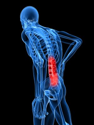
grabbing more grand slam titles, if Nadal’s stem cell treatment is being used to eliminate his pain by repairing his joints or discs, the actual cause of his back pain may not be addressed.

Where does back pain originate?

The number one reason for back pain is muscular and other soft tissue, yet muscles are rarely evaluated as the cause of back pain. The only way to determine if Nadal’s back pain is from soft tissue and similar to most people with back pain would be a physical examination of Nadal’s back that included identifying possible muscles as the cause of his pain.

Some doctors believe that the disc, the cushion between the bones of the spine (the vertebra), is a major cause of back pain. They believe that surgeries to correct the flattening or herniation of the disc will decrease or eliminate back pain. Sometimes they are right, but they are just as likely to be wrong. The truth is that there is as high as a 50% failure rate for spine surgeries that were done to eliminate back pain thought to be related to disc problems. There are other joints in the spine that are thought to cause pain; one of them is the facet joint, which could also be a target for stem cell treatments.

When doctors rely on an MRI or CT scan to determine the source of the pain, the information obtained is often confusing. If a surgeon sees an abnormality on an MRI, he will often point to that abnormality as the cause of the pain; in my experience the abnormality found on an MRI or CT scan frequently is not the cause. In fact, if you randomly selected 100 people off the street, and perform an imaging scan, 40 may present with a herniated disc and have no pain and absolutely no awareness of their herniated disc; 70 may have degenerated (worn) discs with no pain, and a large number will have facet joint abnormalities. Therefore, finding an “abnormality” is more common than not. One, then, can deduce that the abnormality is more likely NOT the source of the pain. So treating the abnormality (with steroid injections, surgery, or stem cells) may therefore not relieve the pain.

Stem Cell Treatment and Sports Stars

Nadal, currently ranked as the number 3 professional tennis player in the world, is not the first sports star to chase after a “miracle cure.” The Denver Bronco quarterback Payton Manning and Yankees pitcher Bartolo Colon both went abroad to seek out stem cell treatment as a quick fix to get back in the game. (They both seem to be doing better overall, but it is inconclusive if the stem cell treatment was the cause of their recovery).

Will it work?

In the laboratory, it has been possible to demonstrate the ability of stem cells (most commonly found in the developing embryo and newborn) to grow new tissue. These cells are like silly putty; they can turn into, or adapt, to become any type of tissue. For example, a stem cell in the right environment in the body could become bone, cartilage or some organ (for example, liver or pancreas). But, it hasn’t been as easy to grow tissue in an actual person. There are some early studies that show that stem cells “may” relieve back pain, but both the doctors who are testing the technique and outside experts say much more research is needed before they can say whether the treatment offers real relief.

The use of stem cell therapies continues to be a hot topic for debate in the sports medicine and orthopedic surgery worlds. There is no current evidence-based research to prove that it works.

Sir William Osler, a famous physician, once said: “Use every new treatment as quickly as possible before it stops working.” Stem cell treatment needs to be further investigated to determine if stem cell treatments indeed work, and if so, for what conditions?


Share
 Page 2 of 7 « 1  2  3  4  5 » ...  Last » 
NORMAN MARCUS PAIN INSTITUTE
30 East 40th Street - New York, NY 10016
Tel 212-532-7999 Fax 212-532-5957
Share
Help Desk Software